Amino acids – Structure of Amino acids
Structure of Amino acids
– Although more than 300 different amino acids have been described in nature, only 20 are commonly found as constituents of mammalian proteins.
– Each amino acid (except for proline, which has a secondary amino group) has a carboxyl group, a primary amino group, and a distinctive side chain (R-group) bonded to the α-carbon atom (Figure 1A).
– At physiologic pH (approximately pH 7.4), the carboxyl group is dissociated, forming the negatively charged carboxylate ion (–COO–), and the amino group is protonated (–NH3+).
– In proteins, almost all of these carboxyl and amino groups are combined through peptide linkage and, in general, are not available for chemical reaction except for hydrogen bond formation (Figure 1B).
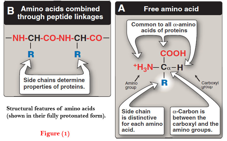
– Thus, it is the nature of the side chains that ultimately dictates the role an amino acid plays in a protein.
– It is, therefore, useful to classify the amino acids according to the properties of their side chains, that is, whether they are nonpolar (have an even distribution of electrons) or polar (have an uneven distribution of electrons, such as acids and bases; Figures 2 and 3).
(1) Amino acids with nonpolar side chains
– Each of these amino acids has a nonpolar side chain that does not gain or lose protons or participate in hydrogen or ionic bonds (Figure 2).
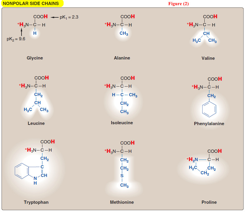
– The side chains of these amino acids can be thought of as (oily) or lipid-like, a property that promotes hydro phobic inter – actions.
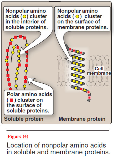
(A) Location of nonpolar amino acids in proteins
– In proteins found in aqueous solutions –a polar environment– the side chains of the nonpolar amino acids tend to cluster together in the interior of the protein (Figure 4).
– This phenomenon, known as the hydrophobic effect, is the result of the hydro phobicity of the nonpolar R-groups, which act much like droplets of oil that coalesce in an aqueous environment.
– The nonpolar R-groups thus fill up the interior of the folded protein and help give it its three-dimensional shape.
– However, for proteins that are located in a hydrophobic environment, such as a membrane, the nonpolar R-groups are found on the outside surface of the protein, interacting with the lipid environment (see Figure 4).
(B) Proline
– Proline differs from other amino acids in that proline’s side chain and α-amino N form a rigid, five-membered ring structure (Figure 5).
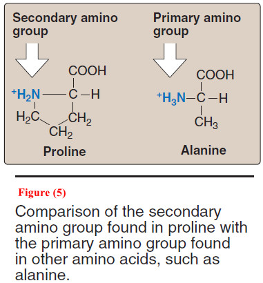
– Proline, then, has a secondary (rather than a primary) amino group. It is frequently referred to as an imino acid.
– The unique geometry of proline contributes to the formation of the fibrous structure of collagen, and often interrupts the α-helices found in globular proteins.
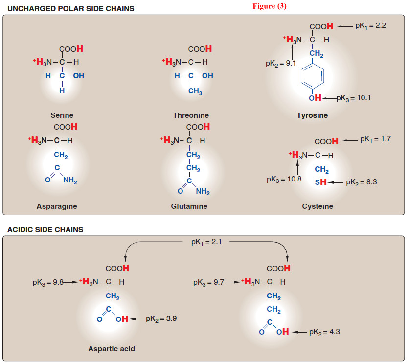
(2) Amino acids with uncharged polar side chains
– These amino acids have zero net charge at neutral pH, although the side chains of cysteine and tyrosine can lose a proton at an alkaline pH (see Figure 3).
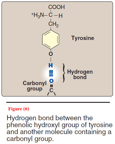
– Serine, threonine, and tyrosine each contain a polar hydroxyl group that can participate in hydrogen bond formation (Figure 6).
– The side chains of asparagine and glutamine each contain a carbonyl group and an amide group, both of which can also participate in hydrogen bonds.
(A) Disulfide bond
– The side chain of cysteine contains a sulf hydryl group (–SH), which is an important component of the active site of many enzymes.
– In proteins, the –SH groups of two cysteines can become oxidized to form a dimer, cystine, which contains a covalent cross-link called a disulfide bond (–S–S–).
(B) Side chains as sites of attachment for other compounds
– The polar hydroxyl group of serine, threonine, and, rarely, tyrosine, can serve as a site of attachment for structures such as a phosphate group.
– In addition, the amide group of asparagine, as well as the hydroxyl group of serine or threonine, can serve as a site of attachment for oligosaccharide chains in glycoproteins.
(3) Amino acids with acidic side chains
– The amino acids aspartic and glutamic acid are proton donors.
– At physiologic pH, the side chains of these amino acids are fully ionized, containing a negatively charged carboxylate group ( COO–).
– They are, therefore, called aspartate or glutamate to emphasize that these amino acids are negatively charged at physiologic pH (see Figure 3).
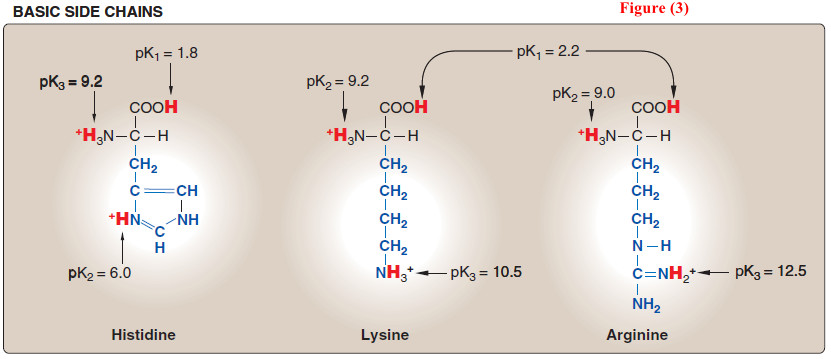
(4) Amino acids with basic side chains
– The side chains of the basic amino acids accept protons (see Figure 3).
– At physiologic pH the side chains of lysine and arginine are fully ionized and positively charged.
– In contrast, histidine is weakly basic, and the free amino acid is largely uncharged at physiologic pH.
– However, when histidine is incorporated into a protein, its side chain can be either positively charged or neutral, depending on the ionic environment provided by the polypeptide chains of the protein.
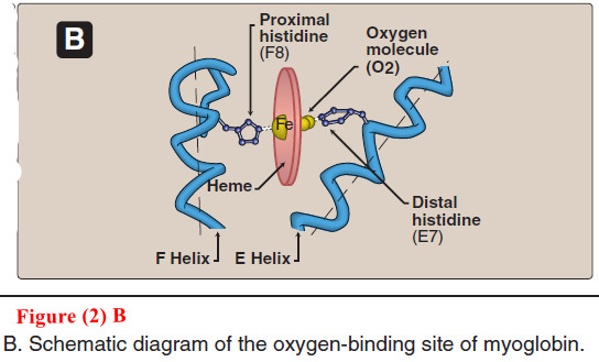
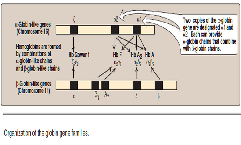
– This is an important property of histidine that contributes to the role it plays in the functioning of proteins such as hemoglobin.
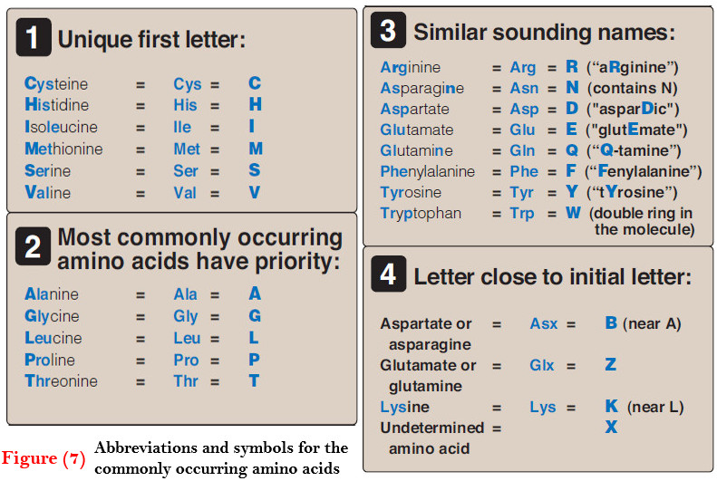
(5) Abbreviations and symbols for commonly occurring amino acids
– Each amino acid name has an associated three-letter abbreviation and a one-letter symbol (Figure 7).
– The one-letter codes are determined by the following rules:
(1) Unique first letter
– If only one amino acid begins with a particular letter, then that letter is used as its symbol.
– For example, I = isoleucine.
(2) Most commonly occurring amino acids have priority
– If more than one amino acid begins with a particular letter, the most common of these amino acids receives this letter as its symbol.
– For example, glycine is more common than glutamate, so G = glycine.
(3) Similar sounding names
– Some one-letter symbols sound like the amino acid they represent.
– For example, F = phenylalanine, or W = tryptophan (“twyptophan” as Elmer Fudd would say).
(4) Letter close to initial letter
– For the remaining amino acids, a oneletter symbol is assigned that is as close in the alphabet as possible to the initial letter of the amino acid, for example, K = lysine.
– Furthermore, B is assigned to Asx, signifying either aspartic acid or asparagine, Z is assigned to Glx, signifying either glutamic acid or glutamine, and X is assigned to an unidentified amino acid.
(6) Optical properties of amino acids
– The α-carbon of an amino acid is attached to four different chemical groups and is, therefore, a chiral or optically active carbon atom
– Glycine is the exception because its α-carbon has two hydrogen substituents and, therefore, is optically inactive.
– Amino acids that have an asymmetric center at the α-carbon can exist in two forms, designated D and L, that are mirror images of each other (Figure 8).
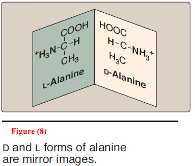
– The two forms in each pair are termed stereoisomers, optical isomers, or enantiomers.
– All amino acids found in proteins are of the L-configuration. However, D-amino acids are found in some antibiotics and in plant and bacterial cell walls.
References:
- Lehninger Principles of Biochemistry / David L. Nelson, Michael M. Cox/ 7th ed, 2017.
- Lippincott’s Illustrated Reviews: Biochemistry / Richard A. Harvey, Denise R. Ferrier/ 5th ed, 2011 / Lippincott Williams & Wilkins, USA.
- Harper’s Illustrated Biochemistry /Robert K. Murray, David A. Bender , Kathleen M. Botham / 28th, 2009/ McGraw-Hill Companies, Inc. USA.