Optical Properties of Sols
Optical Properties of Sols
– In this topic, we will discuss Optical Properties of Sols as follow:
- Sols exhibit Tyndall effect
- Ultramicroscope shows up the presence of individual particles
- Sol particles can be seen with an Electron microscope
(1) Sols exhibit Tyndall effect
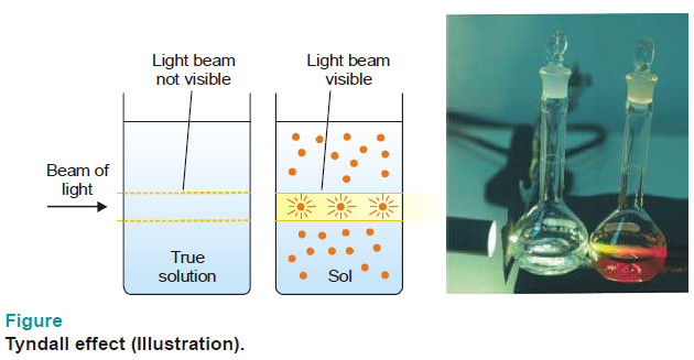
– When a strong beam of light is passed through a sol and viewed at right angles, the path of light shows up as a hazy beam or cone.
– This is due to the fact that sol particles absorb light energy and then emit it in all directions in space.
– This ‘scattering of light’, as it is called, illuminates the path of the beam in the colloidal dispersion.
– The phenomenon of the scattering of light by the sol particles is called Tyndall effect.
– The illuminated beam or cone formed by the scattering of light by the sol particles is often referred as Tyndall beam or Tyndall cone.
– The hazy illumination of the light beam from the film projector in a smoke-filled theatre or the light beams from the headlights of car on a dusty road, are familiar examples of the Tyndall effect.
– If the sol particles are large enough, the sol may even appear turbid in ordinary light as a result of Tyndall scattering.
– True solutions do not show Tyndall effect.
– Since ions or solute molecules are too small to scatter light, the beam of light passing through a true solution is not visible when viewed from the side.
– Thus Tyndall effect can be used to distinguish a colloidal solution from a true solution.
(2) Ultramicroscope shows up the presence of individual particles
– Sol particles cannot be seen with a microscope.
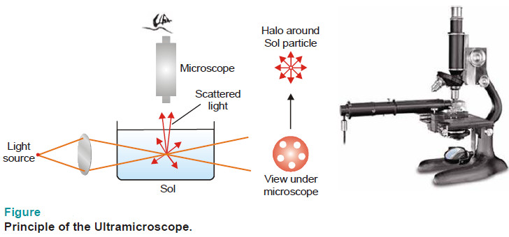
– Zsigmondy (1903) used the Tyndall phenomenon to set up an apparatus named as the ultramicroscope.
– An intense beam of light is focussed on a sol contained in a glass vessel.
– The focus of light is then observed with a microscope at right angles to the beam.
– Individual sol particles appear as bright specks of light against a dark background (dispersion medium).
– It may be noted that under the ultramicroscope, the actual particles are not visible.
– It is the larger halos of scattered light around the particles that are visible.
– Thus an ultramicroscope does not give any information regarding the shape and size of the sol particles.
(3) Sol particles can be seen with an Electron microscope
– In an electron microscope, beam of electrons is focussed by electric and magnetic fields on to a photographic plate.
– This focussed beam is allowed to pass through a film of sol particles.
– Thus it is possible to get a picture of the individual particles showing a magnification of the order of 10,000.
– With the help of this instrument, we can have an idea of the size and shape of several sol particles including paint pigments, viruses, and bacteria.
– These particles have been found to be spheriod, rod-like, disc-like, or long filaments.

